中文
中文 产品细节图:
| 产品编号 | bs-10900R |
| 英文名称 | GAPDH (Loading Control) |
| 中文名称 | 3-磷酸甘油醛脱氢酶(内参)抗体 |
| 别 名 | 38 kDa BFA-dependent ADP-ribosylation substrate; Aging-associated gene 9 protein; BARS-38; cb609; EC 1.2.1.12; G3PD; G3PDH; GAPD; Glyceraldehyde 3 phosphate dehydrogenase;Glyceraldehyde 3 phosphate dehydrogenase liver;Glyceraldehyde 3 phosphate dehydrogenase muscle; KNC-NDS6; MGC102544; MGC102546; MGC103190; MGC103191; MGC105239; MGC127711; MGC88685; OCAS, p38 component; OCT1 coactivator in S phase, 38-KD component; wu:fb33a10. |

|
Specific References (48) | bs-10900R has been referenced in 48 publications.
111 [IF=1.39] Wang, Zhi‑Liang, et al. "Irreversible electroporation‑mediated shRNA knockdown of the HPV18 E6 gene suppresses cervical cancer growth in vitro and in vivo." Oncology Letters. WB ; Human. 222
111 [IF=5.008] Liang et al. Itraconazole exerts its anti-melanoma effect by suppressing Hedgehog, Wnt, and PI3K/mTOR signaling pathways. (2017) Oncotarge. 8:28510-28525 WB ; Human. 222
111 [IF=5.008] Mu et al. Dickkopf-related protein 2 induces G0/G1 arrest and apoptosis through suppressing Wnt/β-catenin signaling and is frequently methylated in breast cancer. (2017) Oncotarge. 8:39443-39459 WB ; Human. 222
111 [IF=4.652] Zong et al. The Effects of Interleukin-17 (IL-17)-Related Inflammatory Cytokines and A20 Regulatory Proteins on Astrocytes in Spinal Cord Cultured In Vitro. (2016) Cell.Physiol.Bioche. 38:1100-10 WB ; Mouse. 222
111 [IF=4.652] Lin et al. MiR-21 Regulates TNF-α-Induced CD40 Expression via the SIRT1-NF-κB Pathway in Renal Inner Medullary Collecting Duct Cells. (2017) Cell.Physiol.Bioche. 41:124-136 WB ; Rat. 222
111 [IF=4.59] Qiao et al. Polydatin Attenuates H2O2-Induced Oxidative Stress via PKC Pathway. (2016) Oxid.Med.Cell.Longe. 2016:5139458 WB ; Human. 222
111 [IF=2.272] Yang et al. Lrig1 is a positive prognostic marker in hepatocellular carcinoma. (2016) Onco.Targets.The. 9:7071-7079 WB ; Human. 222
111 [IF=2.137] Yin et al. Expression and Clinical Significance of ILF2 in Gastric Cancer. (2017) Dis.Marker. 2017:4387081 WB ; Human. 222
111 [IF=1.39] Guo et al. BRAF-activated long non-coding RNA contributes to colorectal cancer migration by inducing epithelial-mesenchymal transition. (2014) Oncol.Let. 8:869-875 WB ; Human. 222
111 [IF=1.39] Wang et al. BRAF-activated long non-coding RNA contributes to cell proliferation and activates autophagy in papillary thyroid carcinoma. (2014) Oncol.Let. 8:1947-1952 WB ; Human. 222
111 [IF=1.238] Liu and Xiao Notch1 signaling induces epithelial-mesenchymal transition in lens epithelium cells during hypoxia. (2017) BMC.Ophthalmo. 17:135 WB ; Human. 222
111 [IF=1.922] Chi et al. Transdermal estrogen gel and oral aspirin combination therapy improves fertility prognosis via the promotion of endometrial receptivity in moderate to severe intrauterine adhesion. (2018) Mol.Med.Rep. 17:6337-6344 WB ; Human. 222
111 [IF=6.34] Zhou et al. Brd4 inhibition attenuates unilateral ureteral obstruction-induced fibrosis by blocking TGF-?-mediated Nox4 expression. (2017) Redox.Biol. 11:390-402 WB ; Rat, Human. 222
111 [IF=1.77] Duan et al. Antidepressant effect of electroacupuncture regulates signal targeting in the brain and increases brain-derived neurotrophic factor levels. (2016) Neural.Regen.Res. 11:1595-1602 WB ; Rat. 222
111 [IF=4.302] Jia et al. Calycosin alleviates allergic contact dermatitis by repairing epithelial tight junctions via down-regulating HIF-1α. (2018) J.Cell.Mol.Med. 22:4507-4521 WB ; Mouse. 222
111 [IF=2.784] Yang et al. miR-1307-3p suppresses the chondrogenic differentiation of human adipose-derived stem cells by targeting BMPR2. (2018) Int.J.Mol.Med. 42:3115-3124 WB ;
111 [IF=2.766] Wan et al. Dietary protein-induced hepatic IGF-1 secretion mediated by PPARγ activation. (2017) PLoS.One. 12:e0173174 WB ; Pig. 222
111 [IF=1.492] Song et al. Extracellular diffusion quantified by magnetic resonance imaging during rat C6 glioma cell progression. (2017) Braz.J.Med.Biol.Res. 50:e5407 WB ; Rat. 222
111 [IF=1.936] Yu B et al. Inhibition of microRNA-143-3p attenuates myocardial hypertrophy by inhibiting inflammatory response. (2018) Nov;42(11):1584-1593. WB ; (SD) Rats. 222
111 [IF=2.976] Guo H et al. Clinical associations between ASCT2 and p-mTOR in the pathogenesis and prognosis of epithelial ovarian cancer. Oncol Rep. 2018 Dec;40(6):3725-3733. WB ; Human. 222
111 [IF=3.448] Liu X et al. Lysosomal dysfunction is associated with persistent lung injury in dams caused by pregnancy exposure to carbon black nanoparticles. Life Sci. 2019 Sep 15;233:116741. WB ; Mouse. 222
111 [IF=2.675] Liu X et al. The lysosomal membrane protein LAMP‐2 is dispensable for PINK1/Parkin‐mediated mitophagy. FEBS Lett. 2019 Nov 6. WB ; Human. 222
111 [IF=2.35] Yu B et al. Suppression of miR-143-3p contributes to the anti-fibrosis effect of atorvastatin on myocardial tissues via the modulation of Smad2 activity. Exp Mol Pathol. 2019 Nov 21;112:104346. WB ; Rat. 222
111 [IF=3.598] Chang L et al. Gypenoside A protects ischemia/reperfusion injuries by suppressing miR‐143‐3p level via the activation of AMPK/Foxo1 pathway. Biofactors. 2019 Dec 30. WB ; Rat. 222
111 [IF=1.851] Wang T et al. The peptide compound urantide regulates collagen metabolism in atherosclerotic rat hearts and inhibits the JAK2/STAT3 pathway. Mol Med Rep. 2020 Mar;21(3):1097-1106. WB ; Rat. 222
111 [IF=4.784] Liu XQ et al. Sodium tanshinone IIA sulfonate protects against Aβ1–42-induced cellular toxicity by modulating Aβ-degrading enzymes in HT22 cells. Int J Biol Macromol. 2020 Feb 6;151:47-55. WB ; Mouse. 222
111 [IF=3.448] Wang T et al. Urotensin receptor antagonist urantide improves atherosclerosis-related kidney injury by inhibiting JAK2/STAT3 signaling pathway in rats. Life Sci. 2020 Feb 13;247:117421. WB ; Rat. 222
111 [IF=.578] Shen D et al. Efficacy evaluation and mechanism study on inhibition of breast cancer cell growth by multimodal targeted fluorescent nanobubbles carrying AMD070 and ICG. Nanotechnology. 2020 Mar 10;31(24):245102. WB ; human. 222
111 [IF=4.268] Wu H et al. A UPLC-Q-TOF/MS-based plasma metabolomics approach reveals the mechanism of Compound Kushen Injection-based intervention against non-small cell lung cancer in Lewis tumor-bearing mice. Phytomedicine . 2020 Jun 2;76:153259. WB ; Mouse. 222
111 [IF=3.647] Lin X et al. Sumoylation enhances the activity of the TGF-β/SMAD and HIF-1 signaling pathways in keloids. Life Sci . 2020 Aug 15;255:117859. WB ; Human. 222
|
| 产品类型 | 内参抗体 |
| 研究领域 | 肿瘤 细胞生物 免疫学 信号转导 新陈代谢 |
| 抗体来源 | Rabbit |
| 克隆类型 | Polyclonal |
| 交叉反应 | Human, Mouse, Rat, |
| 产品应用 |
WB=1:10000-200000 IHC-P=1:100-500 IHC-F=1:100-500 ICC=1:100 IF=1:100-500 (石蜡切片需做抗原修复) not yet tested in other applications. optimal dilutions/concentrations should be determined by the end user. |
| 理论分子量 | 38kDa |
| 细胞定位 | 细胞核 细胞浆 细胞膜 |
| 性 状 | Liquid |
| 浓 度 | 1mg/ml |
| 免 疫 原 | Recombinant human GAPDH full length protein |
| 亚 型 | IgG |
| 纯化方法 | affinity purified by Protein A |
| 缓 冲 液 | 0.01M TBS(pH7.4) with 1% BSA, 0.03% Proclin300 and 50% Glycerol. |
| 保存条件 | Shipped at 4℃. Store at -20 °C for one year. Avoid repeated freeze/thaw cycles. |
| 注意事项 | This product as supplied is intended for research use only, not for use in human, therapeutic or diagnostic applications. |
| PubMed | PubMed |
| 产品介绍 |
Glyceraldehyde 3 phosphate dehydrogenase (GAPDH) is well known as one of the key enzymes involved in glycolysis. As well as functioning as a glycolytic enzyme in cytoplasm, recent evidence suggests that mammalian GAPDH is also involved in a great number of intracellular proceses such as membrane fusion, microtubule bundling, phosphotransferase activity, nuclear RNA export, DNA replication, and DNA repair. During the last decade a lot of data appeared concerning the role of GAPDH in different pathologies including prostate cancer progression, programmed neuronal cell death, age related neuronal diseases, such as Alzheimer's and Huntington's disease. GAPDH is expressed in all cells. It is constitutively expressed in almost all tissues at high levels. There are however some physiological factors such as hypoxia and diabetes that increase GAPDH expression in certain cell types. GAPDH molecule is composed of four 36kDa subunits. Function: Has both glyceraldehyde-3-phosphate dehydrogenase and nitrosylase activities, thereby playing a role in glycolysis and nuclear functions, respectively. Participates in nuclear events including transcription, RNA transport, DNA replication and apoptosis. Nuclear functions are probably due to the nitrosylase activity that mediates cysteine S-nitrosylation of nuclear target proteins such as SIRT1, HDAC2 and PRKDC. Glyceraldehyde-3-phosphate dehydrogenase is a key enzyme in glycolysis that catalyzes the first step of the pathway by converting D-glyceraldehyde 3-phosphate (G3P) into 3-phospho-D-glyceroyl phosphate. Subunit: Homotetramer. Interacts with TPPP; the interaction is direct. Interacts (when S-nitrosylated) with SIAH1; leading to nuclear translocation. Interacts with RILPL1/GOSPEL, leading to prevent the interaction between GAPDH and SIAH1 and prevent nuclear translocation. Interacts with EIF1AD, USP25, PRKCI and WARS. Subcellular Location: Cytoplasm, cytosol. Nucleus. Cytoplasm, perinuclear region. Membrane. Note=Translocates to the nucleus following S-nitrosylation and interaction with SIAH1, which contains a nuclear localization signal. Postnuclear and Perinuclear regions. Post-translational modifications: S-nitrosylation of Cys-152 leads to interaction with SIAH1, followed by translocation to the nucleus. ISGylated (Probable). Sulfhydration at Cys-152 increases catalytic activity. Similarity: Belongs to the glyceraldehyde-3-phosphate dehydrogenase family. SWISS: P04406 Gene ID: 2597 Database links: Entrez Gene: 374193 Chicken Entrez Gene: 2597 Human Entrez Gene: 100042025 Mouse Entrez Gene: 14433 Mouse Entrez Gene: 317743 Zebrafish Omim: 138400 Human SwissProt: P00356 Chicken SwissProt: P04406 Human SwissProt: P16858 Mouse SwissProt: Q5XJ10 Zebrafish GAPDH蛋白几乎在所有组织中都高水平表达,广泛用作Western blot蛋白质标准化的内参,是很好的内参抗体。 GAPDH 作为管家基因在同种细胞或者组织中的蛋白质表达量一般是恒定的。在实验中,可能存在总蛋白浓度测定不准确;或者蛋白质样品在电泳前上样时产生的样品间的操作误差;这些误差需要通过测定每个样品中实际转到膜上的GAPDH的含量来进行校正,所以一般的western实验都需要进行内参设置。具体校正的方法就是将每个样品测得的目的蛋白含量与本样品的GAPDH含量相除,得到每个样品目的蛋白的相对含量。然后才进行样品与样品之间的比较。 甘油醛-3-磷酸脱氢酶(Glyceraldehyde 3 phosphate dehydrogenase,GAPDH)是糖酵解(glycolysis)过程中的关键酶。除了在胞质中作为糖酵解的酶以外,有证据表明哺乳动物细胞中的GAPDH参与了多种胞内生化过程,包括膜融合(membrane fusion)、微管成束(microtubule bundling)、磷酸转移酶(phosphotransferase)激活、核内RNA出核、DNA复制与DNA修复。一些生理因素,诸如低氧(hypoxia)和尿糖(diabetes),可以增加GAPDH在特定细胞中的表达。GAPDH存在于几乎所有的组织中,以高水平持续表达。 GAPDH(甘油醛-3-磷酸脱氢酶)是参与糖酵解的一种关键酶,由4个30-40kDa的亚基组成. |
| 产品图片 |
Sample: 293T(human) cell lysate at 30ug;
Primary: Lane1: Anti-GAPDH (bs-10900R) at 1/2000 dilution Lane2: Anti-GAPDH (bs-10900R) at 1/10000 dilution Lane3: Anti-GAPDH (bs-10900R) at 1/40000 dilution Lane4: Anti-GAPDH (bs-10900R) at 1/80000 dilution Secondary: IRDye800CW Goat Anti-Rabbit IgG at 1/20000 dilution Predicted band size: 38 kD Observed band size: 38kD 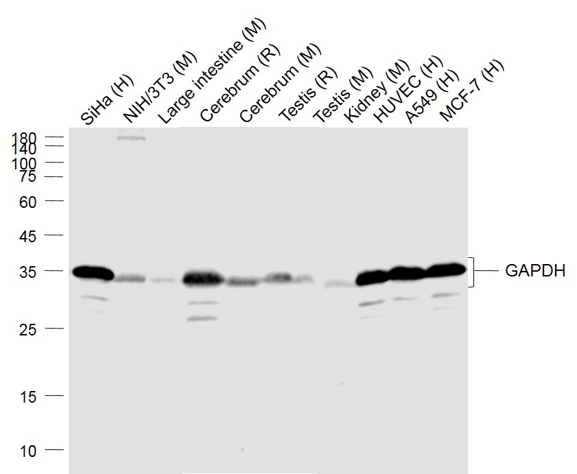
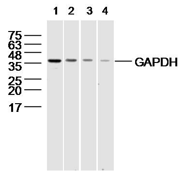
Sample:
Lane 1: SiHa (Human) Cell Lysate at 30 ug Lane 2: NIH/3T3(Mouse) Cell Lysate at 30 ug Lane 3: Large intestine (Mouse) Lysate at 40 ug Lane 4: Cerebrum (Rat) Lysate at 40 ug Lane 5: Cerebrum (Mouse) Lysate at 40 ug Lane 6: Testis (Rat) Lysate at 40 ug Lane 7: Testis (Mouse) Lysate at 40 ug Lane 8: Kidney (Mouse) Lysate at 40 ug Lane 9: HUVEC (Human) Cell Lysate at 30 ug Lane 10: A549 (Human) Cell Lysate at 30 ug Lane 11: MCF-7 (Human) Cell Lysate at 30 ug Primary: Anti-GAPDH (bs-10900R) at 1/1000 dilution Secondary: IRDye800CW Goat Anti-Rabbit IgG at 1/20000 dilution Predicted band size: 36 kD Observed band size: 36 kD 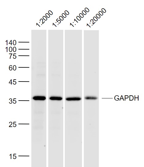
Sample:
293T (Human) Lysate at 40 ug Primary: Anti-GAPDH (bs-10900R) at 1/2000~1/20000 dilution Secondary: IRDye800CW Goat Anti-Rabbit IgG at 1/20000 dilution Predicted band size: 38 kD Observed band size: 36 kD 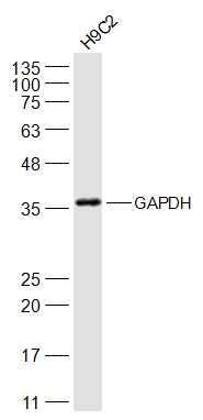
Sample:
H9C2(Rat) Cell Lysate at 30 ug Primary: Anti-GAPDH (bs-10900R) at 1/2000 dilution Secondary: IRDye800CW Goat Anti-Rabbit IgG at 1/20000 dilution Predicted band size: 38 kD Observed band size: 38 kD 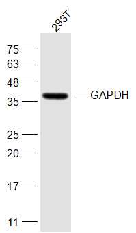
Sample:
293T(Human) Cell Lysate at 30 ug Primary: Anti-GAPDH (bs-10900R) at 1/1000 dilution Secondary: IRDye800CW Goat Anti-Rabbit IgG at 1/20000 dilution Predicted band size: 38 kD Observed band size: 38 kD 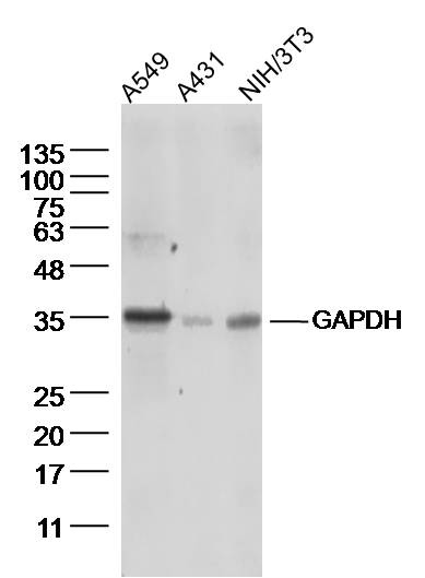
Sample:
A549 Cell (Human) Lysate at 40 ug A431 Cell (Human) Lysate at 40 ug NIH/3T3 Cell (Mouse) Lysate at 40 ug Primary: Anti-GAPDH (bs-10900R) at 1/300 dilution Secondary: IRDye800CW Goat Anti-Rabbit IgG at 1/20000 dilution Predicted band size: 38 kD Observed band size: 36 kD 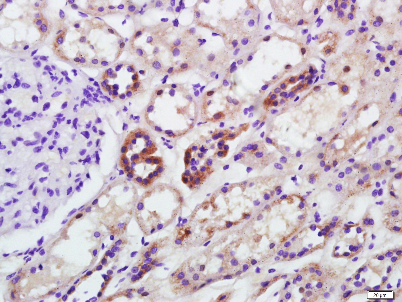
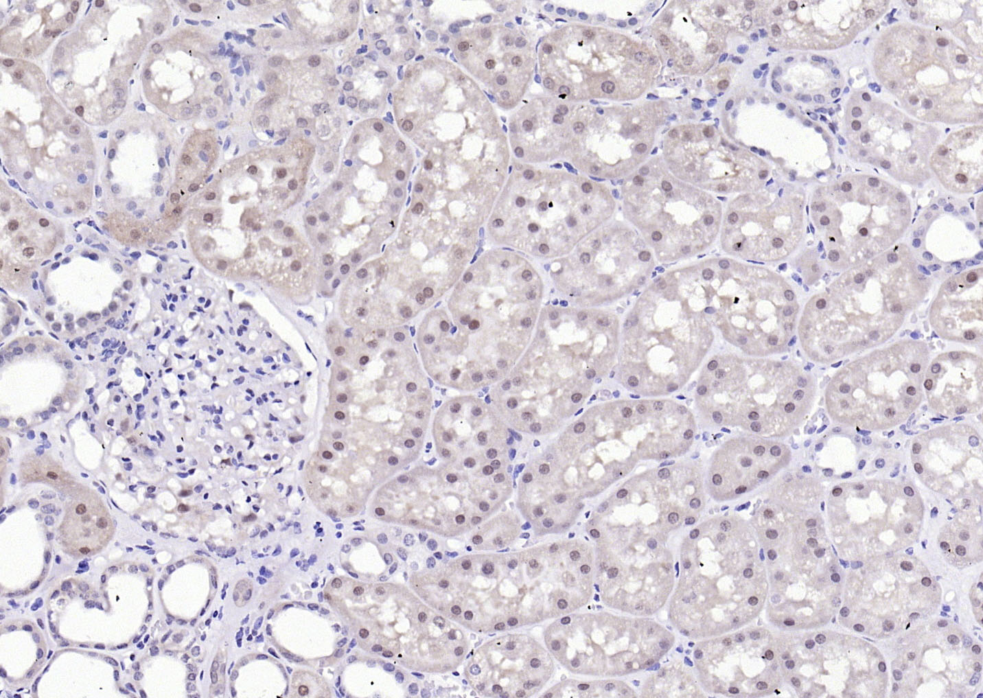
Paraformaldehyde-fixed, paraffin embedded (Human kidney); Antigen retrieval by boiling in sodium citrate buffer (pH6.0) for 15min; Block endogenous peroxidase by 3% hydrogen peroxide for 20 minutes; Blocking buffer (normal goat serum) at 37°C for 30min; Antibody incubation with (GAPDH) Polyclonal Antibody, Unconjugated (bs-10900R) at 1:2000 overnight at 4°C, followed by a conjugated secondary (sp-0023) for 20 minutes and DAB staining.
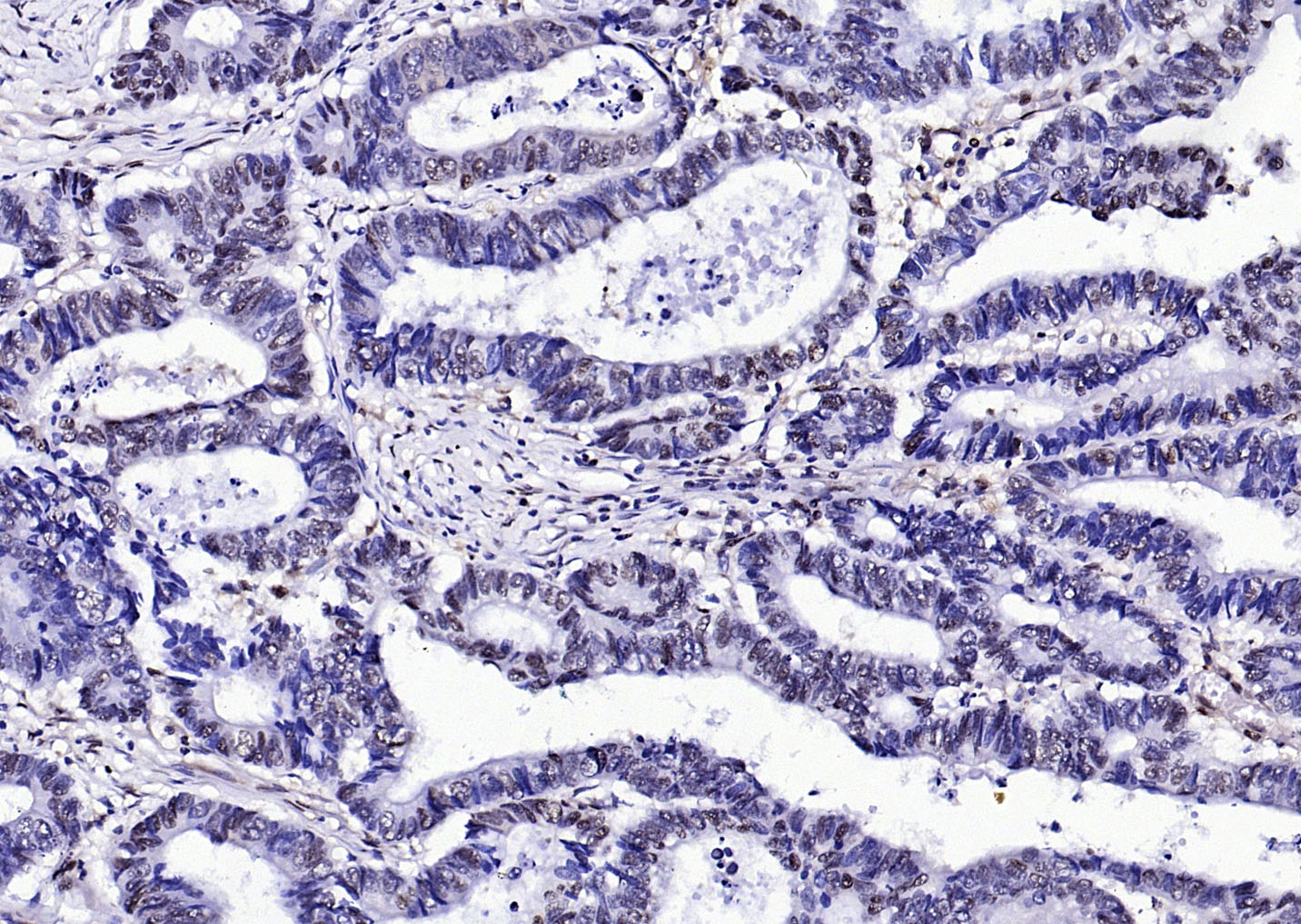
Paraformaldehyde-fixed, paraffin embedded (human colon carcinoma ); Antigen retrieval by boiling in sodium citrate buffer (pH6.0) for 15min; Block endogenous peroxidase by 3% hydrogen peroxide for 20 minutes; Blocking buffer (normal goat serum) at 37°C for 30min; Antibody incubation with (GAPDH (Loading Control)) Polyclonal Antibody, Unconjugated (bs-10900R) at 1:200 overnight at 4°C, followed by operating according to SP Kit(Rabbit) (sp-0023) instructionsand DAB staining.
Paraformaldehyde-fixed, paraffin embedded (Human kidney ); Antigen retrieval by boiling in sodium citrate buffer (pH6.0) for 15min; Block endogenous peroxidase by 3% hydrogen peroxide for 20 minutes; Blocking buffer (normal goat serum) at 37°C for 30min; Antibody incubation with (GAPDH (Loading Control)) Polyclonal Antibody, Unconjugated (bs-10900R) at 1:200 overnight at 4°C, followed by operating according to SP Kit(Rabbit) (sp-0023) instructionsand DAB staining.
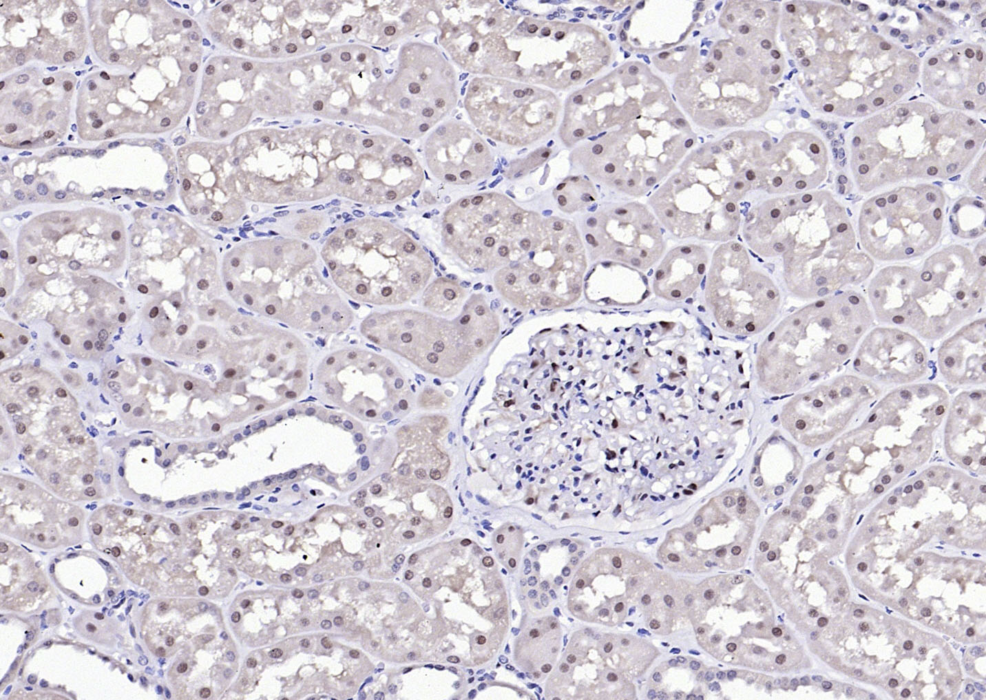
Paraformaldehyde-fixed, paraffin embedded (Human kidney ); Antigen retrieval by boiling in sodium citrate buffer (pH6.0) for 15min; Block endogenous peroxidase by 3% hydrogen peroxide for 20 minutes; Blocking buffer (normal goat serum) at 37°C for 30min; Antibody incubation with (GAPDH (Loading Control)) Polyclonal Antibody, Unconjugated (bs-10900R) at 1:200 overnight at 4°C, followed by operating according to SP Kit(Rabbit) (sp-0023) instructionsand DAB staining.
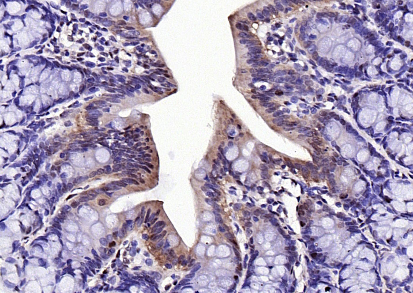
Paraformaldehyde-fixed, paraffin embedded (rat colon); Antigen retrieval by boiling in sodium citrate buffer (pH6.0) for 15min; Block endogenous peroxidase by 3% hydrogen peroxide for 20 minutes; Blocking buffer (normal goat serum) at 37°C for 30min; Antibody incubation with (GAPDH (Loading Control)) Polyclonal Antibody, Unconjugated (bs-10900R) at 1:200 overnight at 4°C, followed by operating according to SP Kit(Rabbit) (sp-0023) instructionsand DAB staining.
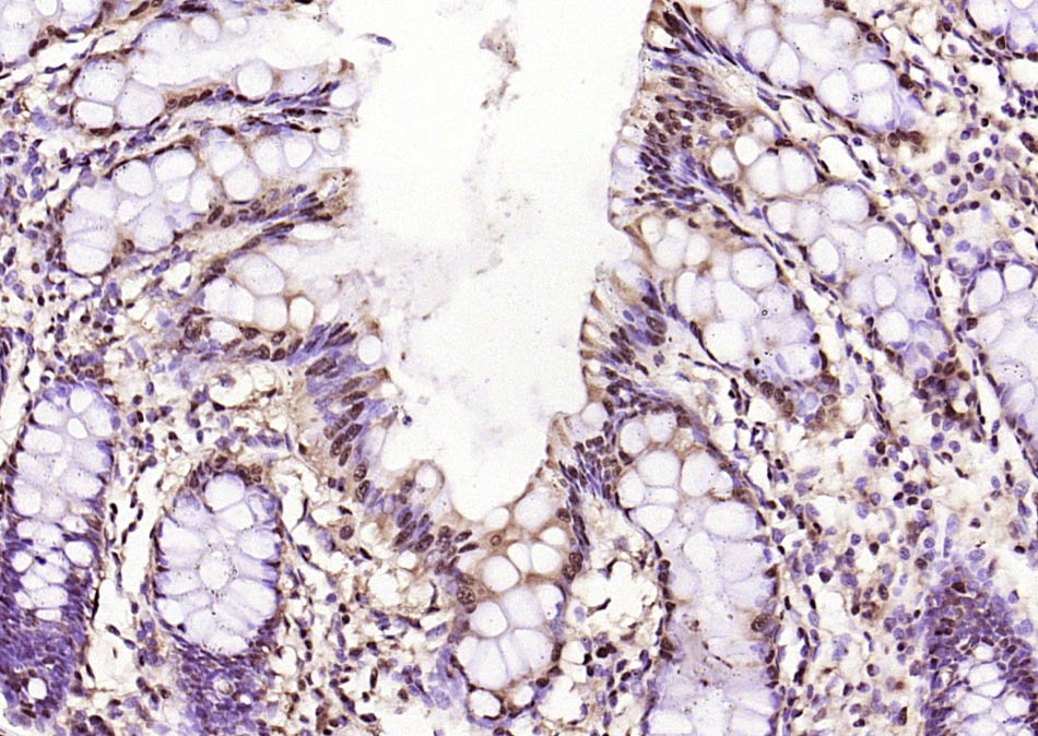
Paraformaldehyde-fixed, paraffin embedded (human colon); Antigen retrieval by boiling in sodium citrate buffer (pH6.0) for 15min; Block endogenous peroxidase by 3% hydrogen peroxide for 20 minutes; Blocking buffer (normal goat serum) at 37°C for 30min; Antibody incubation with (GAPDH (Loading Control)) Polyclonal Antibody, Unconjugated (bs-10900R) at 1:200 overnight at 4°C, followed by operating according to SP Kit(Rabbit) (sp-0023) instructionsand DAB staining.
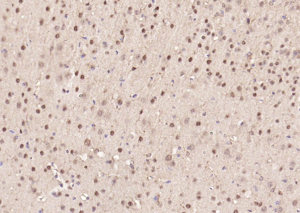
Paraformaldehyde-fixed, paraffin embedded (mouse brain); Antigen retrieval by boiling in sodium citrate buffer (pH6.0) for 15min; Block endogenous peroxidase by 3% hydrogen peroxide for 20 minutes; Blocking buffer (normal goat serum) at 37°C for 30min; Antibody incubation with (GAPDH (Loading Control)) Polyclonal Antibody, Unconjugated (bs-10900R) at 1:200 overnight at 4°C, followed by operating according to SP Kit(Rabbit) (sp-0023) instructionsand DAB staining.
Paraformaldehyde-fixed, paraffin embedded (rat brain); Antigen retrieval by boiling in sodium citrate buffer (pH6.0) for 15min; Block endogenous peroxidase by 3% hydrogen peroxide for 20 minutes; Blocking buffer (normal goat serum) at 37°C for 30min; Antibody incubation with (GAPDH (Loading Control)) Polyclonal Antibody, Unconjugated (bs-10900R) at 1:200 overnight at 4°C, followed by operating according to SP Kit(Rabbit) (sp-0023) instructionsand DAB staining.
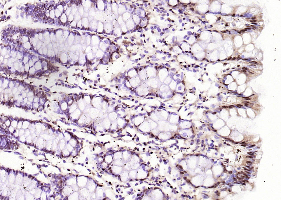
Paraformaldehyde-fixed, paraffin embedded (human colon); Antigen retrieval by boiling in sodium citrate buffer (pH6.0) for 15min; Block endogenous peroxidase by 3% hydrogen peroxide for 20 minutes; Blocking buffer (normal goat serum) at 37°C for 30min; Antibody incubation with (GAPDH (Loading Control)) Polyclonal Antibody, Unconjugated (bs-10900R) at 1:200 overnight at 4°C, followed by operating according to SP Kit(Rabbit) (sp-0023) instructionsand DAB staining.
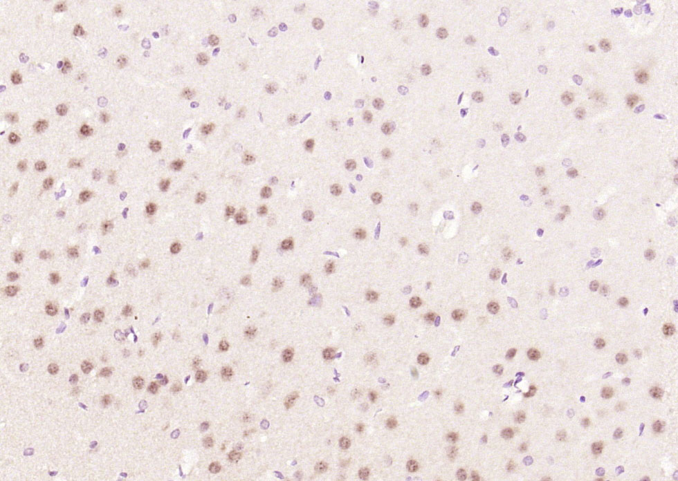
Paraformaldehyde-fixed, paraffin embedded (mouse brain tissue); Antigen retrieval by boiling in sodium citrate buffer (pH6.0) for 15min; Block endogenous peroxidase by 3% hydrogen peroxide for 20 minutes; Blocking buffer (normal goat serum) at 37°C for 30min; Antibody incubation with (GAPDH-Loading Contro) Polyclonal Antibody, Unconjugated (bs-10900R) at 1:400 overnight at 4°C, followed by operating according to SP Kit(Rabbit) (sp-0023) instructionsand DAB staining.
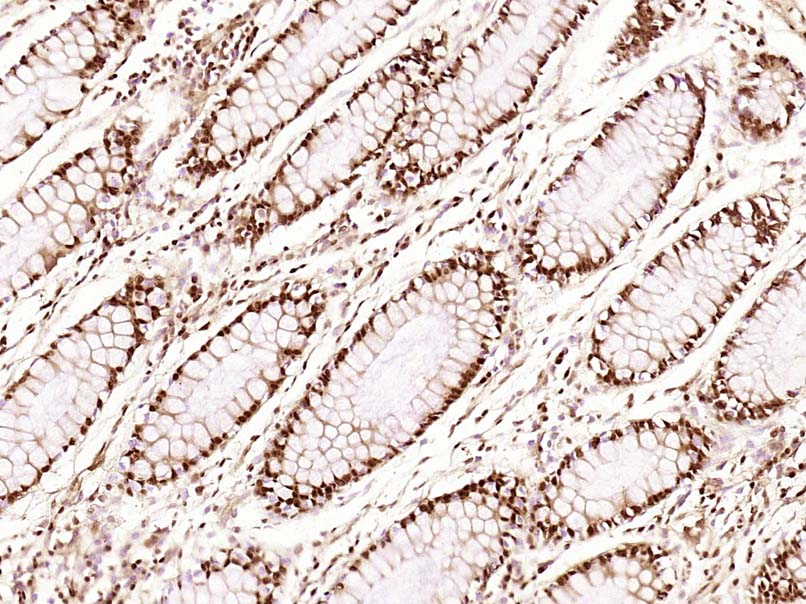
Paraformaldehyde-fixed, paraffin embedded (Human colon carcinoma); Antigen retrieval by boiling in sodium citrate buffer (pH6.0) for 15min; Block endogenous peroxidase by 3% hydrogen peroxide for 20 minutes; Blocking buffer (normal goat serum) at 37°C for 30min; Antibody incubation with (GAPDH) Polyclonal Antibody, Unconjugated (bs-10900R) at 1:500 overnight at 4°C, followed by a conjugated secondary (sp-0023) for 20 minutes and DAB staining.
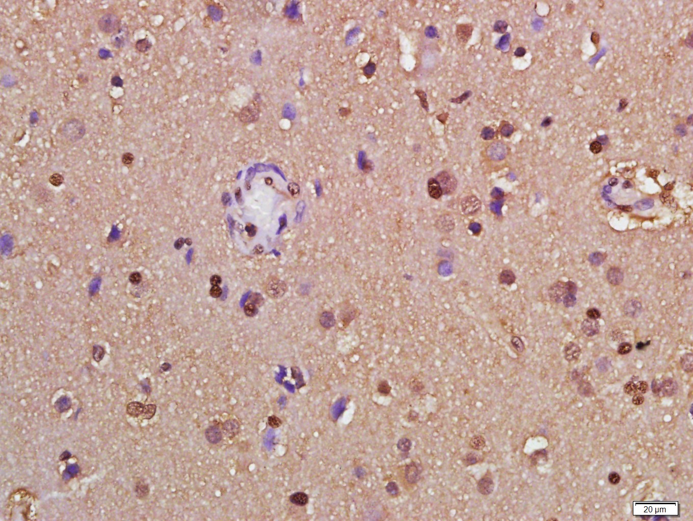
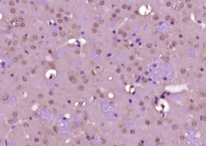
Paraformaldehyde-fixed, paraffin embedded (Human glioma); Antigen retrieval by boiling in sodium citrate buffer (pH6.0) for 15min; Block endogenous peroxidase by 3% hydrogen peroxide for 20 minutes; Blocking buffer (normal goat serum) at 37°C for 30min; Antibody incubation with (GAPDH) Polyclonal Antibody, Unconjugated (bs-10900R) at 1:500 overnight at 4°C, followed by a conjugated secondary (sp-0023) for 20 minutes and DAB staining.
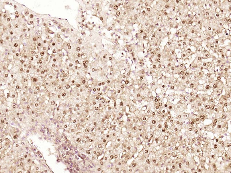
Paraformaldehyde-fixed, paraffin embedded (Human liver cancer); Antigen retrieval by boiling in sodium citrate buffer (pH6.0) for 15min; Block endogenous peroxidase by 3% hydrogen peroxide for 20 minutes; Blocking buffer (normal goat serum) at 37°C for 30min; Antibody incubation with (GAPDH) Polyclonal Antibody, Unconjugated (bs-10900R) at 1:500 overnight at 4°C, followed by a conjugated secondary (sp-0023) for 20 minutes and DAB staining.
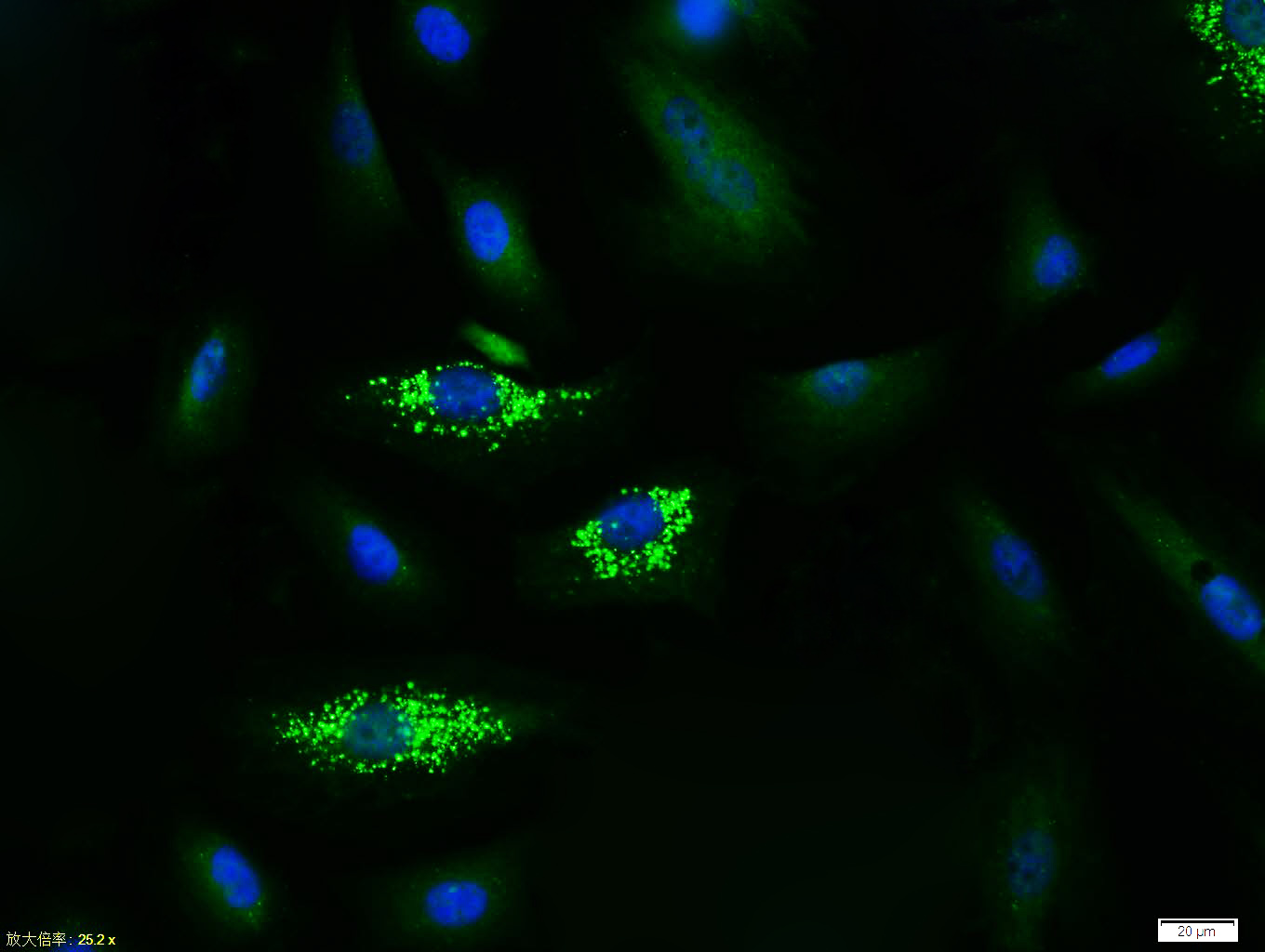
Tissue/cell: A549 cell; 4% Paraformaldehyde-fixed; Triton X-100 at room temperature for 20 min; Blocking buffer (normal goat serum, C-0005) at 37°C for 20 min; Antibody incubation with (GAPDH (Loading Control)) polyclonal Antibody, Unconjugated (bs-10900R) 1:100, 90 minutes at 37°C; followed by a FITC conjugated Goat Anti-Rabbit IgG antibody at 37°C for 90 minutes, DAPI (blue, C02-04002) was used to stain the cell nuclei.

Tissue/cell: A549 cell; 4% Paraformaldehyde-fixed; Triton X-100 at room temperature for 20 min; Blocking buffer (normal goat serum, C-0005) at 37°C for 20 min; Antibody incubation with (GAPDH (Loading Control)) polyclonal Antibody, Unconjugated (bs-10900R) 1:100, 90 minutes at 37°C; followed by a FITC conjugated Goat Anti-Rabbit IgG antibody at 37°C for 90 minutes, DAPI (blue, C02-04002) was used to stain the cell nuclei.
|
 暂时没有说明书
1.《Multifunction Sr, Co and F co-doped microporous coating on titanium of antibacterial, angiogenic and osteogenic activities》
作者:Jianhong Zhou ,Lingzhou Zhao 影响因子:4.259
期刊:《Scientific Reports 6, Article number: 29069 (2016)》 PMID:27353337
作者:Jianhong Zhou ,Lingzhou Zhao 影响因子:4.259
期刊:《Scientific Reports 6, Article number: 29069 (2016)》 PMID:27353337
| 用户名: | (可为空) |
| E-mail: | *(必填) |
| 评价等级: |


|
| 评论内容: |
*(必填)
|